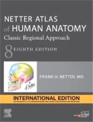
醫學領域必備圖譜!!!
對於正在學習解剖學、參與臨床解剖、或與患者說明解剖知識的學生和臨床專業人士來說,《Netter Atlas of Human Anatomy 8E》以臨床醫生的視角,依區域詳細的說明人體。同時強調對臨床醫師在培訓和實作中最重要的解剖關係的插圖。內含超過550張精緻的圖表以及幾十張精心挑選的常見放射學影像。
Key Features
•Presents world-renowned, superbly clear views of the human body from a clinical perspective, with paintings by Dr. Frank Netter as well as Dr. Carlos A. G. Machado, one of today’s foremost medical illustrators.
•Content guided by expert anatomists and educators: R. Shane Tubbs, Paul E. Neumann, Jennifer K. Brueckner-Collins, Martha Johnson Gdowski, Virginia T. Lyons, Peter J. Ward, Todd M. Hoagland, Brion Benninger, and an international Advisory Board.
•Offers region-by-region coverage, including muscle table appendices at the end of each section and quick reference notes on structures with high clinical significance in common clinical scenarios.
•Contains new illustrations by Dr. Machado including clinically important areas such as the pelvic cavity, temporal and infratemporal fossae, nasal turbinates, and more.
•Features new nerve tables devoted to the cranial nerves and the nerves of the cervical, brachial, and lumbosacral plexuses.
•Uses updated terminology based on the second edition of the international anatomic standard, Terminologia Anatomica, and includes common clinically used eponyms.
•Enhanced eBook version included with purchase. Your enhanced eBook allows you to access all of the text, figures, and references from the book on a variety of devices.
•Provides access to extensive digital content: every plate in the Atlas and over 100 bonus plates including illustrations from previous editions is enhanced with an interactive label quiz option and supplemented with "Plate Pearls" that provide quick key points of the major themes of each plate. Digital content also includes over 300 multiple choice questions and other learning tools.
| FindBook |
有 1 項符合
Netter Atlas of Human Anatomy: Classic Regional Approach, 8th Edition (International Edition)的圖書 |
 |
Netter Atlas of Human Anatomy: Classic Regional Approach (IE) 作者:Netter 出版社:Elsevier Science Health Science div 出版日期:2022-04-01 規格:28.5cm*22.2cm (高/寬) / 8 / 平裝 / 676頁 |
| 圖書館借閱 |
| 國家圖書館 | 全國圖書書目資訊網 | 國立公共資訊圖書館 | 電子書服務平台 | MetaCat 跨館整合查詢 |
| 臺北市立圖書館 | 新北市立圖書館 | 基隆市公共圖書館 | 桃園市立圖書館 | 新竹縣公共圖書館 |
| 苗栗縣立圖書館 | 臺中市立圖書館 | 彰化縣公共圖書館 | 南投縣文化局 | 雲林縣公共圖書館 |
| 嘉義縣圖書館 | 臺南市立圖書館 | 高雄市立圖書館 | 屏東縣公共圖書館 | 宜蘭縣公共圖書館 |
| 花蓮縣文化局 | 臺東縣文化處 |
|
|
圖書介紹 - 資料來源:博客來 評分:
圖書名稱:Netter Atlas of Human Anatomy: Classic Regional Approach, 8th Edition (International Edition)
|


